April 11, 2023
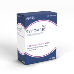
The buzz in the retina community is the potential of finally having a treatment for geographic atrophy (GA), the dry, atrophic form of macular degeneration. It is expected that by the first half of 2023 we will have not just one, but possibly two treatments for GA. Phase 3 data on pegcetacoplan by Apellis Pharmaceuticals and avacincaptad pegol by Iveric Bio have been submitted to the FDA for review. In November 2022, the FDA granted breakthrough therapy status for avacincaptad pegol. Meanwhile, the U.S. Food and Drug Administration has approved SYFOVRE (pegcetacoplan injection) for the treatment of GA secondary to age-related macular degeneration (AMD).
AMD is considered to be the leading cause of legal blindness in adults over the age of 65, affecting approximately 10 million people in the U.S. and over 186 million worldwide. This number is expected to increase to 288 million by 2040.1 Approximately 85% to 90% of patients have the dry form of the disease, while 10% to 15% progress to wet, neovascular AMD (nAMD).2
Geographic atrophy represents the late stage of dry AMD and affects approximately one million people in the U.S. and up to five to eight million worldwide. It is responsible for 10% to 20% of cases of legal blindness. Though GA is considered to be the least common form of AMD, it may be more common than we realize. In the AREDS studies, the 10-year risk of progression from intermediate AMD to GA was 53.9% compared to progression to nAMD, which was 47.6%.3 In a 2015 meta-analysis of late stage AMD among whites, the annual incidence rate of GA was 1.9 per 1,000 Caucasians aged ≥ 50; slightly less than the incidence of nAMD, which was 1.8 per 1,000 Caucasians.4
ECPs Need to Do Better When Detecting Geographic Atrophy
The problem is we have not been focused on GA detection, but rather conversion to nAMD, so GA gets missed or overlooked, especially smaller lesions. Our exams traditionally are geared around the detection of AMD — are there drusen or retinal pigment epithelial (RPE) mottling? These are features common in dry AMD and easily seen on traditional funduscopy, often becoming an indication for performing imaging studies such as fundus photos, optical coherence tomography/optical coherence tomography angiography (OCT/OCTA), or fundus autofluorescence (FAF). Once AMD is detected, the question then becomes whether there is any fluid or subretinal hemorrhage, all of which are markers of nAMD. The fact is, small areas of GA may be present, but it goes unrecognized or even ignored unless it becomes large enough or a threat to central vision. Indeed, the image we usually conjure in our mind of GA is that of a large atrophic lesion that is either threatening central fixation or already has.
The reality is GA starts small and usually well outside of the fovea. Unless you look carefully and/or know what to look for, early GA can be missed or overlooked. Fundus photos are not the best imaging modality to detect early GA simply because early lesions can be small and there is poor contrast between the “normal” retina and areas of RPE atrophy. Instead, OCT or FAF are much better at detecting even small areas of atrophy.
Autofluoresence is usually the best at detecting GA as it easily highlights areas of hypofluoresence, a hallmark FAF feature of GA. OCT is not quite as good, but once you know what to look for can be just as sensitive as FAF. Both the B-scan and the en face image can highlight areas of GA. When looking at a B-scan image of GA, a hallmark feature is a transmission defect through the RPE into the choroid. The en face image does a great job of defining the boundaries of the GA. Both FAF and the en face OCT image can also be used to measure the lesion size as well as measuring the change in lesion size over time. Most commercial OCTs have integrated software that can do this automatically. As AMD patients return for follow-up exams, reviewing previous OCT or FAF images with a focus on GA detection will often show areas of GA that are present but may not have been recognized. In addition, as we compare repeat scans over time, we can also see the lesions slowly increasing in size.
With two potential treatments for GA, all eye care providers will need to do a better job of GA detection. Optometry in particular is uniquely positioned being on the front lines of primary eye care. Many of these patients will be diagnosed by an optometrist. With growing adoption of diagnostic retinal imaging technologies, we have all the tools needed for early diagnosis and management. Then we can refer for treatment when appropriate.
Studies Determine Effectiveness of Both Treatments
How effective are these potentially new treatments for GA? Both pegcetacoplan and avacincaptad pegalol are complement inhibitors but work in different areas of the complement pathway. Pegcetacoplan is a C3 inhibitor, whereas avacincaptad pegol blocks C5. Both drugs shut down activation of inflammatory processes and cell-killing membrane attack complex. Both medications are delivered as a monthly intravitreal injection.
GATHER1 and GATHER2 were independent Phase 3 studies comparing avacincaptad pegol vs. a sham injection. Both studies met the prespecified one-year primary end points with GATHER1 showing a 35.4% reduction in GA growth vs. the sham and GATHER2 showing a 17.7% reduction in GA growth vs. sham.5 With these positive results, avacincaptad pegol became the first drug to deliver two Phase 3 studies that met their pre-specified primary end points at 12 months of slowing GA progression.
Pegcetacoplan also delivered positive Phase 3 results at 12 months but only in one of the Phase 3 trials, the DERBY. OAKS and DERBY compared monthly and every other month (EOM) injection of pegacetacoplan vs. sham. OAKS showed statistically significant reductions in GA lesion growth vs sham in the monthly and EOM arms by 22% and 16%. DERBY did not reach statistical significance: pegcetacoplan decreased GA lesion growth vs. sham by 12% and 11% in the monthly and EOM arms, respectively.6 However, it is important to note that the treatment results were much better as the study continued beyond 12 months. At 18 and 24 months, an accelerated treatment effect in both studies was seen compared to the sham. At 24 months, DERBY showed a 36% monthly, 29% EOM reduction in GA progression, whereas OAKS had 24% monthly, 25% EOM reduction in slowing GA progression.7 For these reasons, Apellis waited to submit the 18- to 24-month data to the FDA, instead of only submitting the 12-month data.
Finally, There Is Hope for Patients!
Clearly, there is a treatment benefit with both drugs, but it’s important to recognize it’s not a cure. It’s also important to recognize these are patients who will need to be treated every month, and it may take up to a year of monthly injections to achieve any meaningful benefit. If patients can get beyond a year, we can really see the therapeutic benefit accelerate. Remember, though, the majority of GA patients will have good acuity and may not be as motivated to commit to an every-month treatment regimen when they aren’t experiencing an immediate visual benefit.
As providers on the front lines of AMD, it’s not unreasonable to question the therapeutic efficacy of these drugs that need to be given every month in order to see a 15% to 30% slower growth rate of GA over a one-year period. Is it worth it? Is that good enough? If we take a broader view and look beyond a year, perhaps it is. For the patient who has already lost central vision in one eye due to GA, there is no doubt it will be worth it.
There are a lot of questions that will need to be answered. When should these patients be referred for treatment? Is there a certain size of GA or proximity to the fovea that would indicate when patients should be referred and/or treated? No doubt, as these medications become approved and we gain more experience, many of these questions will be answered. In the meantime, we think of all the elderly patients we have had in our exam chairs with GA slowly losing central vision, as well as their independence, as we helplessly watch, impotent at being able to stop or slow it down. Finally, there is hope!
References
1 Wong WL, Su X, Li X, et al. Global prevalence of age-related macular degeneration and disease burden projection for 2020 and 2040: a systematic review and meta-analysis. Lancet Glob Health. 2014;2(2):e106-116
2 accessed January 11, 2023. What is Macular Degeneration?
3 Chew EY, Clemons, TE, Agron E, et al. Ten-Year Follow-up of Age-Related Macular Degeneration in the Age-Related Eye Disease StudyAREDS Report No. 36. JAMA Ophthalmol. 2014;132(3):272-277.
4 Rudnicka AR, Kapetanakis VV, Jarrar Z, et al. Incidence of late-stage age-related macular degeneration in American whites: systematic review and meta-analysis. Am J Ophthalmol. 2015;160(1):85-93.
5 Khanani AM, Patel SS, Staurenghi G. et al. The Efficacy of Avacincaptad Pegol in Geographic Atrophy: GATHER1 and GATHER2 Results. Retina Society 2022. https://ivericmedical.com/static/RetinaSociety2022_Efficacy-f610417058de3027f34fd055737ebc79.pdf
6 Apellis announces pegcetacoplan showed continuous and clinically meaningful effects at month 18 in phase 3 DERBY and OAKS studies for geographic atrophy (GA). https://investors.apellis.com/news-releases/news-release-details/apellis-announces-pegcetacoplan-showed-continuous-and-clinically. Accessed January 11, 2023
7 Apellis Announces 24-Month Results Showing Increased Effects Over Time with Pegcetacoplan in Phase 3 DERBY and OAKS Studies in Geographic Atrophy (GA) https://investors.apellis.com/news-releases/news-release-details/apellis-announces-24-month-results-showing-increased-effects


